TruQuick™ D-Dimer
The TruQuick D-Dimer (Whole Blood/ Plasma) is a rapid chromatographic immunoassay for the qualitative detection of human D-dimer in whole blood or plasma as an aid in the diagnosis of Disseminated Intravascular Coagulopathy (DIC), deep venous thrombosis (DVT) and pulmonary embolism (PE).
D-dimer (or D dimer) is a fibrin degradation product (or FDP), a small protein fragment present in the blood after a blood clot is degraded by fibrinolysis. It is so named because it contains two crosslinked D fragments of the fibrin protein.1D-dimer concentration may be determined by a blood test to help diagnose thrombosis. Since its introduction in the 1990s, it has become an important test performed in patients with suspected thrombotic disorders. While a negative result practically rules out thrombosis, a positive result can indicate thrombosis but does not rule out other potential causes. Its main use, therefore, is to exclude thromboembolic disease where the probability is low. In addition, it is used in the diagnosis of the disorder Disseminated Intravascular Coagulopathy.1 The TruQuick D-Dimer (Whole Blood/ Plasma) is a simple test that utilizes a combination of anti- D-dimer antibody coated particles and capture reagents to qualitatively detect D-dimer in whole blood or plasma. The minimum detection level is 500ng/mL.
The TruQuick D-Dimer (Whole Blood/ Plasma) is a qualitative, membrane based immunoassay for the detection of D-dimer in whole blood or plasma. The membrane is pre-coated with specific capture antibodies in the test line regions of the test. During testing, the whole blood or plasma specimen reacts with the particle coated with specific antibodies. The mixture migrates upward on the membrane chromatographically by capillary action to react with specific capture antibodies on the membrane and generate a colored line. The presence of this colored line in the specific test line region indicates a positive result, while its absence indicates a negative result. To serve as a procedural control, a colored line will always appear in the control line region indicating that proper volume of specimen has been added and membrane wicking has occurred.
- Bring the pouch to room temperature before opening it. Remove the test cassette from the sealed pouch and use it within one hour.
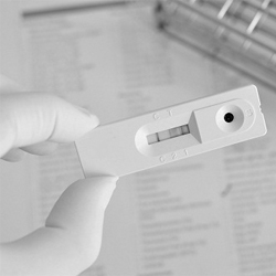
- Place the cassette on a clean and level surface.
- For Plasma specimen:
- Hold the dropper vertically and transfer 1 drop of plasma (approximately 25L) to the specimen area, then add 2 drops of buffer (approximately 80L), and start the timer. See illustration below.
- For Venipuncture Whole Blood specimen:
- Hold the dropper vertically and transfer 1 drop of whole blood (approximately 25 L) to the specimen area, then add 2 drops of buffer (approximately 80 L), and start the timer. See illustration below.
- For Fingerstick Whole Blood specimen:
- To use a capillary tube: Fill the capillary tube and transfer approximately 25 L of fingerstick whole blood specimen to the specimen area of test cassette, then add 2 drops of buffer (approximately 80 L) and start the timer. See illustration below.
- To use hanging drops: Allow 1 hanging drop of fingerstick whole blood specimen (approximately 25 L) to fall into the specimen area of test cassette, then add 2 drops of buffer (approximately 80 L) and start the timer. See illustration below.
- For Plasma specimen:
- Wait for the colored line(s) to appear. Read results at 10 minutes. Do not interpret the result after 20 minutes.