TruQuick™ Rota/Adeno Combo
Acute diarrhea disease in young children is a major cause of morbidity worldwide and is a leading cause of mortality in developing countries.1 Rotavirus is the most common agent responsible for acute gastroenteritis, mainly in young children.2 Its discovery in 1973 and its association with infantile gastroenteritis represented a very important advancement in the study of gastroenteritis not caused by acute bacterial infection. Rotavirus is transmitted by oral-fecal route with an incubation period of 1-3 days. Although specimens collected within the second and fifth day of the illness are ideal for antigen detection, the rotavirus may still be found while diarrhea continues. Rotaviral gastroenteritis may result in mortality for populations at risk such as infants, the elderly and immunocompromised patients.3 In temperate climates, rotavirus infections occur mainly in the winter months. Endemics as well as epidemics affecting some thousand people have been reported.4 With hospitalized children suffering from acute enteric disease, up to 50% of the analyzed specimen are positive for rotavirus.5 The viruses replicate in the cell nucleus and tend to be host species specific, producing a characteristic cytopathic effect (CPE). Because rotavirus is extremely difficult to culture, it is unusual to use isolation of the virus in diagnosing an infection. Instead, a variety of techniques have been developed to detect rotavirus in feces.
- To collect fecal specimens: Collect sufficient quantity of feces (1-2 mL or 1-2 g) in a clean, dry specimen collection container to obtain enough virus particles. Best results will be obtained if the assay is performed within 6 hours after collection. Specimen collected may be stored for 3 days at 2-8 C if not tested within 6 hours. For long-term storage, specimens should be kept below – 20 C.
- To process fecal specimens:
- For Solid Specimens: Unscrew the cap of the Specimen Collection Tube, then randomly stab the specimen collection applicator into the fecal specimen in at least 3 different sites to collect approximately 50 mg of feces (equivalent to 1/4 of a pea). Do not scoop the fecal specimen.
- For Liquid Specimens: Hold the dropper vertically, aspirate fecal specimens, and then transfer 2 drops of the liquid specimen (approximately 50 μL) into the Specimen Collection Tube containing the Extraction Buffer. Tighten the cap onto the Specimen Collection Tube, then shake the Specimen Collection Tube vigorously to mix the specimen and the Extraction Buffer. Leave the collection tube sit for 2 minutes.
- Bring the pouch to room temperature before opening it. Remove the Test Cassette from the foil pouch and use it within one hour. Best results will be obtained if the test is performed immediately after opening the foil pouch.
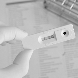
- Hold the Specimen Collection Tube upright and open the cap on the tip. Invert the Specimen Collection Tube and transfer 2 full drops of the extracted specimen (approximately 80 μL) to the specimen well (S) of the Test Cassette, then start the timer. Avoid trapping air bubbles in the specimen well (S). See illustration below.
- Read the results at 10 minutes after dispensing the specimen. Do not read results after 20 minutes.
- Wadell, G. Laboratory diagnosis of infectious diseases: Principles and Practices. New York: Springer-Verlag. 1988;(2):284-300.
- Wilhelmi I, Roman E, Sanchez-Fauquier A. Viruses causing gastroenteritis. Clin Microbiol Infect. 2003 April(9):247-262.
- Cubitt, WD. Rotavirus infection: An unexpected hazard in units caring for the elderly. Geriatric Medicine Today. 1982;1:33-38.
- Hung, T et al. Waterborne outbreak of rotavirus diarrhoea in adults in china caused by a novel rotavirus. Lancet. 1984 May 26;1:8387:1139-1142.
- Cukor G, Perron DM, Hudson R, Blacklow NR. Detection of rotavirus in human stools by using monoclonal antibody. J Clin Microl. 1984;19:888-892.
- Wood DJ, Bailey AS. Detection of adenovirus types 40 and 41 in stool specimens by immune electron microscopy. J Med Virol. 1987;21:191-199.
- Osamu N, et al. Enzyme-linked immunosorbent assay employing monoclonal antibodies for direct identification of enteric adenoviruses (Ad40, 41) in feces. Microbiol Immunol. 1990;34(10):871-877.
- Wood DJ, Bijlsma K, de Jong JC, Tonkin C. Evaluation of a commercial monoclonal antibody-based enzyme immunoassay for detection of adenovirus types 40 and 41 in stool specimens. J Clin Microbiol. 1989; June27(6):1155-1158.
- Thomas E, et al. The utility of latex agglutination assays in the diagnosis of pediatric viral gastroenteritis. Am J Clin Pathol. 1994;101:742-746.