IgG Rheumatoid Factor Mouse ELISA
Rheumatoid factors are autoantibodies against Fc region of of IgG and is found in 70-80% of patients suffering from chronic rheumatoid arthritis, and are considered to be closely related to its pathological syndrome. Experimental animal models with spontaneous autoimmune diseases similar to those in humans, and animals with artificially-induced inflammation have been used to elucidate the mechanism of autoimmune diseases and to search for potential new remedies. A representative model animal of spontaneous autoimmune diseases is MRL/lpr mouse. As MRL/lpr shows high incidence of lymph node tumor, nephritis, angitis, and arthritis, this animal strain is useful for studies of the mechanism of human autoimmune diseases including rheumatoid arthritis.
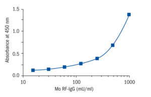
Autoantibodies found in MRL/lpr serum are IgG type rheumatoid factor, IgM type rheumatoid factor, anti-ssDNA antibodies, anti-dsDNA antibodies, and anti-Sm antibody, etc. This kit enables quantification and comparison of IgG type rheumatoid factor with a calibration curve using standard antibody preparation
Research topic
Animal studies
Type
Direct ELISA, HRP-labelled antibody
Applications
Serum, Plasma-EDTA, Plasma-Citrate
Sample Requirements
1 µl/well
Storage/Expiration
Store the complete kit at 2–8°C. Under these conditions, the kit is stable until the expiration date (see label on the box).
Calibration Curve

Calibration Range
15.6–1 000 mU/ml
Limit of Detection
0.78 mU/ml
– Ishihara K, Sawa S, Ikushima H, Hirota S, Atsumi T, Kamimura D, Park SJ, Murakami M, Kitamura Y, Iwakura Y, Hirano T. The point mutation of tyrosine 759 of the IL-6 family cytokine receptor gp130 synergizes with HTLV-1 pX in promoting rheumatoid arthritis-like arthritis. Int Immunol. 2004 Mar;16 (3):455-65
– Kikukawa T, Kojima M, Abe C. A novel assay kits for autoantibodies rate on spontaneous autoimmune model mice. Jap J Inflammation. 2000;20:697-701
– Kumanogoh A, Shikina T, Watanabe C, Takegahara N, Suzuki K, Yamamoto M, Takamatsu H, Prasad DV, Mizui M, Toyofuku T, Tamura M, Watanabe D, Parnes JR, Kikutani H. Requirement for CD100-CD72 interactions in fine-tuning of B-cell antigen receptor signaling and homeostatic maintenance of the B-cell compartment. Int Immunol. 2005 Oct;17 (10):1277-82
– Oishi H, Mizuki S, Terada M, Kudo M, Araki K, Araki M, Nose M, Takahashi S. Increased expression of soluble form of vascular cell adhesion molecule-1 aggravates autoimmune arthritis in MRL-Fas(lpr) mice. Pathol Int. 2007 Nov;57 (11):734-40
– Takeda Y, Takeno M, Iwasaki M, Kobayashi H, Kirino Y, Ueda A, Nagahama K, Aoki I, Ishigatsubo Y. Chemical induction of HO-1 suppresses lupus nephritis by reducing local iNOS expression and synthesis of anti-dsDNA antibody. Clin Exp Immunol. 2004 Nov;138 (2):237-44
– Yim YK, Lee H, Hong KE, Kim YI, Lee BR, Son CG, Kim JE. Electro-acupuncture at acupoint ST36 reduces inflammation and regulates immune activity in Collagen-Induced Arthritic Mice. Evid Based Complement Alternat. 2007 Mar;4 (1):51-7

