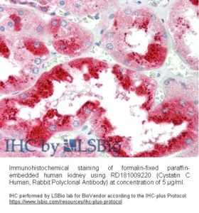
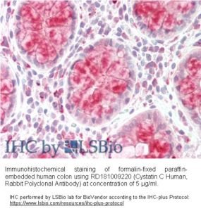
Cystatin C Human, Rabbit Polyclonal Antibody
Cystatin C is a non-glycosilated basic single-chain protein consisting of 120 amino acids with a molecular weight of 13.36 kDa and is characterized by two disulfide bonds in the carboxy-terminal region. It belongs to the cystatins superfamilly which inactivates lysosomal cysteine proteinases, e.g. cathepsin B, H,.K, L and S. Imbalance between Cystatin C and cysteine proteinases is associated with inflammation, renal failure, cancer, Alzheimer’s disease, multiple sclerosis and hereditary Cystatin C amyloid angiopathy. Its increased level has been found in patients with autoimune diseases, with colorectal tumors and in patients on dyalisis. Serum Cystatin C seems to be better marker of glomerular filtration rate than creatinine. On the other hand, low concentration of Cystatin C presents a risk factor for secondary cardiovascular events.
Type
Polyclonal Antibody
Applications
Western blotting, ELISA, Immunohistochemistry
Antibodies Applications


Source of Antigen
Human urine
Hosts
Rabbit
Preparation
The antibody was raised in rabbits by immunization with the Human Cystatin C.
Species Reactivity
Human
Purification Method
Immunoaffinity chromatography on a column with immobilized Human Cystatin C.
Antibody Content
0.1 mg (determined by BCA method, BSA was used as a standard)
Formulation
The antibody is lyophilized in 0.05 M phosphate buffer, 0.1 M NaCl, pH 7.2. AZIDE FREE.
Reconstitution
Add 0.1 ml of deionized water and let the lyophilized pellet dissolve completely. Slight turbidity may occur after reconstitution, which does not affect activity of the antibody. In this case clarify the solution by centrifugation.
Shipping
At ambient temperature. Upon receipt, store the product at the temperature recommended below.
Storage/Expiration
The lyophilized antibody remains stable and fully active until the expiry date when stored at –20°C. Aliquot the product after reconstitution to avoid repeated freezing/thawing cycles and store frozen at –80°C. Reconstituted antibody can be stored at 4°C for a limited period of time; it does not show decline in activity after one week at 4°C.
Quality Control Test
Indirect ELISA – to determine titer of the antibody
SDS PAGE – to determine purity of the antibody
Note
This product is for research use only. This product will be replaced with RD181009220–01
– Danjo A, Yamaza T, Kido MA, Shimohira D, Tsukuba T, Kagiya T, Yamashita Y, Nishijima K, Masuko S, Goto M, Tanaka T. Cystatin C stimulates the differentiation of mouse osteoblastic cells and bone formation. Biochem Biophys Res Commun . Aug 17;360(1):199-204 (2007)
– Konduri SD, Yanamandra N, Siddique K, Joseph A, Dinh DH, Olivero WC, Gujrati M, Kouraklis G, Swaroop A, Kyritsis AP, Rao JS. Modulation of cystatin C expression impairs the invasive and tumorigenic potential of human glioblastoma cells. Oncogene . Dec 12;21(57):8705-12 (2002)
– Nedelkov D, Nelson RW. Delineating protein-protein interactions via biomolecular interaction analysis-mass spectrometry. J Mol Recognit . Jan-Feb;16(1):9-14 (2003)
– Nedelkov D, Nelson RW. Design and use of multi-affinity surfaces in biomolecular interaction analysis-mass spectrometry (BIA/MS): a step toward the design of SPR/MS arrays. J Mol Recognit . Jan-Feb;16(1):15-9 (2003)

