C-Peptide (U-type) Mouse ELISA
Insulin is first synthesized as a single chain polypeptide, proinsulin, then three disulfide bonds are formed, and finally divided into insulin and C-peptide through enzymatic splitting. Mouse C-peptide 1 is a single chain peptide composed of 29 amino acids, while C-peptide 2 is composed of 31 residues. C-peptide is secreted together with insulin. The role of C-peptide has been considered to keep the best configuration to form three disulfide bonds, and has no biological activity, however, recent studies revealed that C-peptide can bind, probably, a G-proteincoupling specific receptor present on the surface of endothelial cells, kidney microtubule cells and fibroblasts, resulting in activation of calcium-dependent intracellular signaling, activation of Na+-K+-ATPase, and enhancement of NO synthesis. Administration of C-peptide to DM1 patients enhances blood circulation in the skeletal muscle and skin, and also minimizes kidney glomerular hyperfiltration, decreasing albumin excretion into urine, and also improves nervous function, indicating that C-peptide should be given together with insulin to DM1 patients. Important region to bind receptor has been reported to be C-terminal pentapeptide (27-31).
The biological half life of C-peptide is several times longer than that of insulin. Measurement of C-peptide is useful in estimation of pancreatic function for insulin synthesis and secretion. Urinary C-peptide concentration is well correlated to its blood level. C-peptide measurement is also useful in estimation of insulin secretion by cultured islet of Langerhans because very often insulin is added to the culture medium, and it is difficult to discriminate secreted insulin from added insulin. As Shibayagi’s kit recognizes the common sequences between C-peptide 1 and 2, it can measure total amount of C-peptide.
Research topic
Animal studies, Diabetology – Insulin, C-Peptide, Proinsulin
Type
Sandwich ELISA, Biotin-labelled antibody
Applications
Serum, Plasma-EDTA, Plasma-Heparin, Plasma-Citrate, Cell culture medium
Sample Requirements
10 µl/well
Storage/Expiration
Store the complete kit at 2–8°C. Under these conditions, the kit is stable until the expiration date (see label on the box).
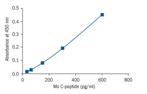
Calibration Curve

Calibration Range
30–3 000 pg/ml
Limit of Detection
1.5 pg/ml
– Guo W, Miao C, Liu S, Qiu Z, Li J, Duan E. Efficient differentiation of insulin-producing cells from skin-derived stem cells. Cell Prolif. 2009 Feb;42 (1):49-62
– Hori Y, Fukumoto M, Kuroda Y. Enrichment of putative pancreatic progenitor cells from mice by sorting for prominin1 (CD133) and platelet-derived growth factor receptor beta. Stem Cells. 2008 Nov;26 (11):2912-20
– Ibii T, Shimada H, Miura S, Fukuma E, Sato H, Iwata H. Possibility of insulin-producing cells derived from mouse embryonic stem cells for diabetes treatment. J Biosci Bioeng. 2007 Feb;103 (2):140-6
– Liu MJ, Shin S, Li N, Shigihara T, Lee YS, Yoon JW, Jun HS. Prolonged remission of diabetes by regeneration of beta cells in diabetic mice treated with recombinant adenoviral vector expressing glucagon-like peptide-1. Mol Ther. 2007 Jan;15 (1):86-93
– Narushima M, Kobayashi N, Okitsu T, Tanaka Y, Li SA, Chen Y, Miki A, Tanaka K, Nakaji S, Takei K, Gutierrez AS, Rivas-Carrillo JD, Navarro-Alvarez N, Jun HS, Westerman KA, Noguchi H, Lakey JR, Leboulch P, Tanaka N, Yoon JW. A human beta-cell line for transplantation therapy to control type 1 diabetes. Nat Biotechnol. 2005 Oct;23 (10):1274-82
– Torisu T, Sato N, Yoshiga D, Kobayashi T, Yoshioka T, Mori H, Iida M, Yoshimura A. The dual function of hepatic SOCS3 in insulin resistance in vivo. Genes Cells. 2007 Feb;12 (2):143-54

