TruQuick™ Calprotectin
TruQuick Calprotectin is a rapid chromatographic immunoassay for the qualitative detection of Calprotectin in feces specimens. It is to aid in the diagnosis of inflammatory gastrointestinal disorders.
Calprotectin is a 24 kDa dimer of calcium-binding proteins S100A8 and S100A9.1 The complex accounts for up to 60% of the soluble protein content of the neutrophil cytosol.2 Calprotectin becomes available in the intestinal lumen via leukocyte shedding,3 active secretion,2 cell disturbance, and cell death.3 This results in elevated fecal calprotectin levels, which can be detected in the stool.3 Elevated fecal calprotectin levels can indicate migration of neutrophils into the intestinal mucosa, which occurs during intestinal inflammation.4 Fecal calprotectin has been used to detect intestinal inflammation, and can serve as a marker for inflammatory bowel diseases.5 Calprotectin is useful as a marker, as it is resistant to enzymatic degradation, and can be easily measured in feces.6
- To collect fecal specimens: Collect sufficient quantity of feces (1-2 mL or 1-2 g) in a clean, dry specimen collection container to obtain maximum antigens (if present). Best results will be obtained if the assay is performed within 6 hours after collection. Specimens may be stored for 3 days at 2-8 C if not tested within 6 hours. For long term storage, specimens should be kept below -20 C.
- To process fecal specimens:
- For Solid Specimens:
- Unscrew the cap of the Specimen Collection Tube, then randomly stab the specimen collection applicator into the fecal specimen in at least 3 different sites to collect approximately 50 mg of feces (equivalent to 1/4 of a pea). Do not scoop the fecal specimen.
- For Liquid Specimens:
- Hold the dropper vertically, aspirate fecal specimens, and then transfer 2 drops (approximately 80 μL) into the Specimen Collection Tube containing the Extraction Buffer.
- For Solid Specimens:
- Tighten the cap onto the Specimen Collection Tube, then shake the tube vigorously to mix the specimen and the Extraction Buffer. Leave the tube alone for 2 minutes.
- Bring the pouch to room temperature before opening it. Remove the Test Cassette from the foil pouch and use it within one hour. Best results will be obtained if the test is performed immediately after opening the foil pouch.
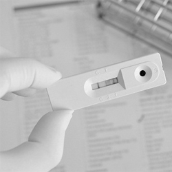
- Hold the Specimen Collection Tube upright and open the cap onto the Specimen Collection Tube. Invert the tube and transfer 2 full drops of the extracted specimen (approximately 80 μL) to the specimen well (S) of the Test Cassette, then start the timer. Avoid trapping air bubbles in the specimen well (S). See illustration below.
- Read results at 5 minutes after dispensing the specimen. Do not read results after 10 minutes.
- Note: If the specimen does not migrate (presence of particles), centrifuge the extracted specimens contained in the Extraction Buffer vial. Collect 80 μL of supernatant, dispense into the specimen well (S) of a new Test Cassette and start afresh following the instructions mentioned above.
- Brophy MB, Nolan EM. Manganese and microbial pathogenesis: Sequestration by the mammalian immune system and utilization by microorganisms. ACS Chem Biology. 2015 Jan 16;10:150.
- Striz I, Trebichavsky I. Calprotectin – a pleiotropic molecule in acute and chronic inflammation.. Physiological research / Academia Scientiarum Bohemoslovaca. 2004;53 (3):245–53.
- Lehmann FS, Burri E, Beglinger C. The role and utility of faecal markers in inflammatory bowel disease. Therapeutic Advances in Gastroenterology. 2014 Oct 13;8 (1): 23–36.
- Gupta R. Biomarkers in toxicology. San Diego, CA: Academic Press. 2014;272–273.
- Marshall WM, Lapsley M, Day A, Ayling R. Clinical biochemistry: Metabolic and clinical aspects (3 ed.). Elsevier Health Sciences. 2014.
- Tibble J, Teahon K, Thjodleifsson B, Roseth A, Sigthorsson G, Bridger S, et al. A simple method for assessing intestinal inflammation in Crohn’s disease. Gut. 2000;47 (4):506-13.